
The Spinal Fusion Surgery Process: What Patients Can Expect
Spinal fusion surgery is a commonly performed procedure designed to stabilize the spine and reduce pain by permanently connecting two or more vertebrae. While the idea of “fusing” bones may sound complex, the process is highly structured and carefully planned to ensure safety and optimal outcomes.
Understanding each stage—from preparation to recovery—can help patients feel more informed and confident about their care.
What Is Spinal Fusion Surgery?
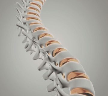
Spinal fusion is a surgical technique that joins two or more vertebrae into a single, solid bone. By eliminating motion between these bones, the procedure can relieve pain, improve stability, and correct spinal deformities.
It is often recommended for conditions such as:
- Degenerative disc disease
- Spinal instability or fractures
- Herniated discs
- Scoliosis or other deformities
- Spinal stenosis
Step 1: Pre-Surgical Preparation
Before surgery, patients undergo a thorough evaluation to ensure they are good candidates for the procedure. This phase typically includes:
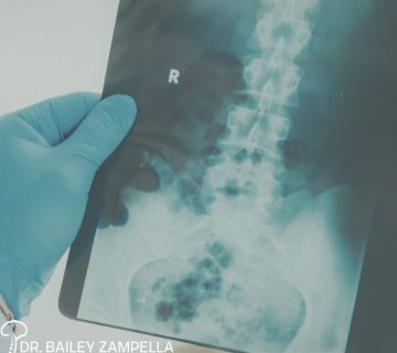
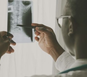
- Physical examination and imaging (MRI, CT, or X-rays)
- Blood tests and medical clearance
- Medication review and adjustments
- Pre-operative instructions (such as fasting or stopping certain medications)
Planning ahead for post-surgical care—like arranging transportation and home support—is also essential.
Step 2: Anesthesia and Surgical Setup
On the day of surgery, patients are placed under general anesthesia, meaning they are fully asleep and pain-free during the procedure.
The surgical team monitors vital signs closely throughout the operation, ensuring patient safety at every step.
Step 3: Accessing the Spine
The surgeon begins by making an incision to reach the affected area of the spine. Depending on the condition being treated, this approach may be:
- From the back (posterior approach)
- From the front (anterior approach)
- From the side (lateral approach)
The choice depends on the location of the problem and the patient’s anatomy.
Step 4: Preparing the Bone Graft
A key component of spinal fusion is the bone graft, which helps the vertebrae grow together. This graft may come from:

- The patient’s own body (often the pelvis)
- A donor (bone bank)
- Synthetic or bioengineered materials
The graft acts as a bridge, encouraging new bone growth between the vertebrae.
Step 5: Fusing the Vertebrae
Once the graft is prepared, the surgeon places it between the targeted vertebrae. To hold everything in place, medical hardware such as screws, rods, or plates may be used. ()
Over time, the bone graft and vertebrae heal together, forming a single solid structure—much like a natural bone healing process.
Step 6: Closing the Incision
After the fusion is complete, the surgeon closes the incision using sutures or staples. The patient is then moved to a recovery area, where medical staff monitor them as they wake from anesthesia.
Step 7: Recovery and Rehabilitation
Recovery from spinal fusion surgery is gradual and varies by patient. Key aspects include:
- Hospital stay: Typically 2–3 days
- Pain management: Medication and monitoring
- Activity restrictions: Limited bending, lifting, or twisting
- Physical therapy: Helps restore strength and mobility
- Healing time: Several months for the bones to fully fuse
Patients may also be advised to wear a brace to support the spine during healing.
What Results Can Patients Expect?
Spinal fusion can significantly reduce pain and improve spinal stability. However, outcomes vary depending on the underlying condition and overall health. While the fused segment no longer moves, many patients experience improved quality of life and function.
